Before we get into the development of the urinary system, we should understand what is the embryo trying to build??

The urinary system function to remove the waste products from the body. It helps to maintain a normal concentration of water and electrolytes in the body fluids, regulates pH and volume of body fluids, helps control the production of RBCs and blood pressure.
The urinary system consists of two bean shaped kidneys that remove the waste products from blood and form urine, two tubular ureters which transport urine from the kidneys to the sac-like urinary bladder. Urine is stored in the urinary bladder and then conveyed to the outside by the urethra.
The kidneys are located in the on each side of the vertebral column between the 12th thoracic vertebrae and the forth lumber vertebrae. With a convex lateral surface and a concave medial surface; blood vessels, nerves, lymphatic vessels and the ureter leaves and enter the kidneys from the kidney’s hilum. The superior end of the ureter expands to form a funnel-shaped area called the renal pelvis. The pelvis is subdivided into two or three tubes called major calyces (singular, calyx), which in turn are further subdivided into several minor calyces.
Each kidney has two distinctive layers: an inner medulla and an outer granulated cortex. The renal medulla is composed of conical masses of tissue called the renal pyramids. The outer cortex surrounds the medulla and dips into it between the renal pyramids forming the renal columns (Figure 1).
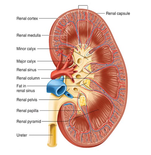
Figure 1: Longitudinal section of a kidney showing the major stuructures. (Shier et al, 2006)
The Nephron
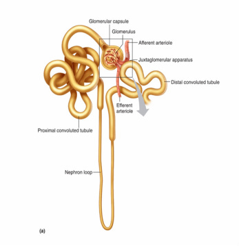
Figure 2: Structure of a nephron. (shier et al, 2006)
The functional unit of the kidney is the nephron. A nephron consists of a renal corpuscle and a renal tubule. A renal corpuscle is composed of a glomerulus which filter the blood, and a glomerular capsule which receives the filtrate and lead away to form the proximal convoluted tubule. The proximal convoluted tubule dips down towards the renal pelvis forming the descending limb of the nephron loop and then curves up again from the ascending limb of the nephron loop. The ascending loop then coils tightly to form the distal convoluted tubule. The distal convoluted tubule from several nephrons merge to form the collecting duct which in turn enlarges as more distal convoluted tubule merge in. the collecting duct open into the minor calyx (figure 2).
The kidneys are supplied by renal arties, lateral branches of the abdominal aorta, which arise between the first and the second lumbar vertebrae. Blood is drained by renal veins which pass anterior to the renal arteries and join the inferior vena cava. Lymph is drained to lateral aortic nodes around the origin of the renal arteries.
The kidneys are supplied by renal arties, lateral branches of the abdominal aorta, which arise between the first and the second lumbar vertebrae. Blood is drained by renal veins which pass anterior to the renal arteries and join the inferior vena cava. Lymph is drained to lateral aortic nodes around the origin of the renal arteries.
