In a cranial to caudal sequence, three sets of excretory organs develop in human embryos: pronephrons, mesonephrons and metanephrons.PronephrosEarly in the fourth week, pronephros represented by 7-10 cell clusters appear in the neck region. By the end of the fourth week, all indications of the pronephric system are degenerated (figure 3).
|
 |
Mesonephros
During the degeneration of the pronephros in the fourth week, the first excretory tubules of the mesonephros start to appear. They lengthen rapidly and acquire a tuft of capillaries that form a glomerulus at their medial extremity, tubules around the glomerulus form Bowman’s capsule, and together those structures constitute the renal corpuscle. Laterally, the tubules enter the longitudinal collecting duct known as the mesonephric or walffian duct (figure 3).
The urogenital ridge is formed in the middle of the second month as a large ovoid organ on each side of the midline. Cranial tubules and glomeruli show degenerative changes while caudal ones are still differentiating; however, by the second month, the majority disappears. In males, few caudal tubules remain and take part in the formation of the genital system.
The urogenital ridge is formed in the middle of the second month as a large ovoid organ on each side of the midline. Cranial tubules and glomeruli show degenerative changes while caudal ones are still differentiating; however, by the second month, the majority disappears. In males, few caudal tubules remain and take part in the formation of the genital system.
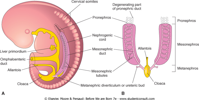
Figure 3: Excretory organs in an embryo during the fifth week. A, Lateral view; B, Ventral view. (Moore and Persaud, 2008)
Metanephros
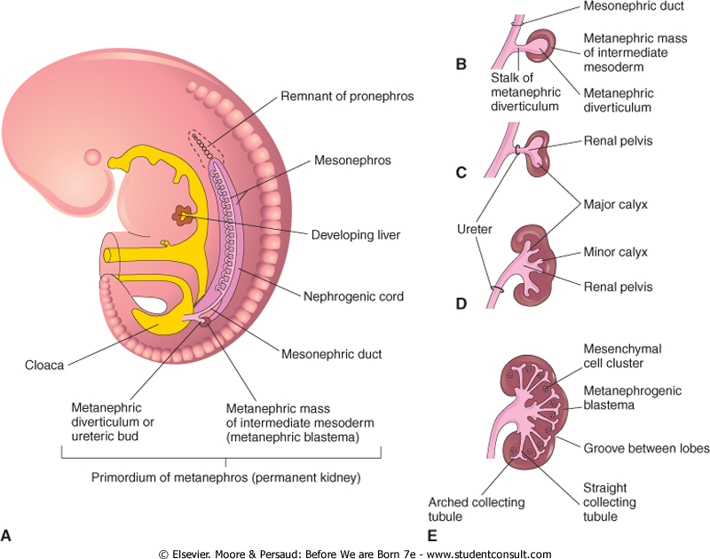
Figure 4: A, development of the metanephrons, lateral view; B to E, stages in teh development of the metanephric diverticulum. (Moore and Persaud,2008)
Metanephros, or the definitive kidneys, begin to develop early in the fifth week and start to function approximately 4 weeks later. The definitive kidneys develop from two sources:
1. The metanephric diverticulum (ureteric bud), which is the primordium of the ureter, the renal pelvic, the calices and the collecting tubules.
2.The metanephric mass of the intermediate mesoderm (metanephric blastema), which would form the nephrons.
The metanephric diverticulum is an outgrowth from the mesonephric duct. It elongates penetrating the metanephrogenic blastema. The stalk of the metanephrephric diverticulum forms the ureter, its cranial end forms the renal pelvis, and the branching bud would form the collecting tubules. The straight collecting tubules branch and subdivide to form generations of collecting tubules. The first four generations form the major calices, the second four generation form the minor calices and the remaining generations form the collecting tubules (figure 4).
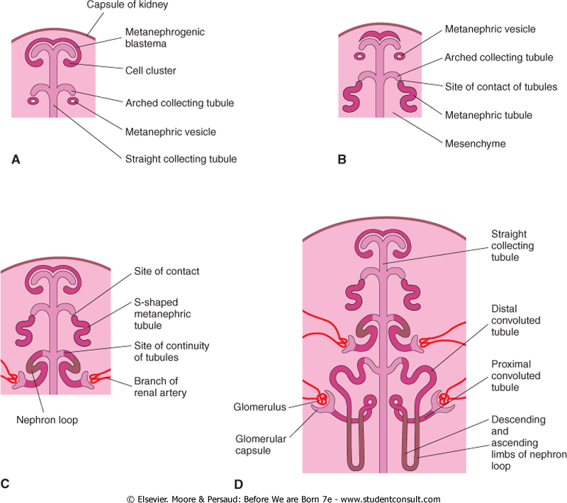
Figure 5: Stages of the development of nephrons. (Moore and Persaud, 2008)
The end of the collecting tubules induces the metanephric mass to form metanephric vesicles. These vesicles elongate and give rise to S-shaped tubule. Capillaries grow at one end of the S-shaped tubules and differentiate into glomeruli. The glomeruli and the tubules form the nephrons. Bowmans capsule, which is indented by the glomeruli, forms at the proximal end of the developing nephron. The distal end forms an open connection with one of the collecting tubules. Continuous lengthening of the S-shaped tubules give rise to the proximal convoluted tubules, loop of Henle, and the distal convoluted tubule (figure 5).
The definitive kidney becomes functional by the 12th week. Urine is passed to the amniotic cavity and is mixed by the amniotic fluid. During foetal life, the kidneys are not responsible for the excretion of waste products. The kidneys usually have lobulated appearance soon after birth; however, this appearance disappears as nephrons mature and grow.
The molecular regulation of the development of the kidneys involves many transcriptional factors interactions. The mesenchyme expresses WT1 which enables the tissue to respond to induction by the ureteric bud. The mesenchyme also produce glial derived neurotropic factor (GDNF) and hepatocyte growth factor (HGF). Both of which are regulated by WT1 and stimulate the branching and growth of the ureteric bud. Meanwhile, the bud produce Fibroblast growth factor 2 (FGF2) and bone morphogenetic protein 7 (BMP7) which stimulate proliferation in the metanephric mesenchyme and maintain expression of WT1. Branches of the ureteric bud secret WNT9B and WNT6 which upregulate PAX2 and WNT4 in the metanephric mesenchyme. PAX2 and WNT4, promotes condensation of the mesenchyme and causes the condensed mesenchyme to epithelialize and form tubules respectively. In addition, extracellular matrix changes and is replaced with laminin and type IV collagen forming the basement membrane of the epithelial cells.
 |
 |
